Research
- Research Overview
- Organo Metallic Chemical Vapor Deposition
- Optical Response of Gold Nanoparticle Clusters
- Evanescent Wave Analytical Tools
- Surface Functionalisation
- Detection of Optically Active Molecules
- Optical Tweezers in Evanescent Fields
- Evanescent Microscopy and Spectroscopy
- Research Opportunities
- Research Group
- Former Coworkers and Students
- Collaborators
- Publications
Contact Information
Prof. Silvia Mittler
Physics & Astronomy 240
(519) 661-2111 x.88592
smittler@uwo.ca
FAX: (519) 661-2033
Waveguide Evanescent Field (WEFF), Waveguide Evanesevent Field Scattering (WEFS) Microscopy and Wavguide Absorption Spectroscopy
The evanescent field of a propagating waveguide mode can be used in various ways in microscopy and spectroscopy. It basically serves as the illumination source for samples located on the surface of a waveguide.
The evanescent light as the illuminating source of any microscopic object on the surface of the waveguide can serve many purposes. It can be used for:
- scattering light microscopy, where a contast arises from low and high scattering areas
- fluorescence microscopy, where the evanescent field excites chromophores
- absorption phenomena
The objects may vary from inorganic and organic particles or patterned surfaces to biological objects like cells and bacteria. The wavelength of illumination can easily be chosen, as well as the intensity. To some extend the depth profile can be adjusted by applying the right waveguide mode. Another option is to define a destinct polarisation state by using either TE or TM modes or a combination of both.
Evanescent Microscopy
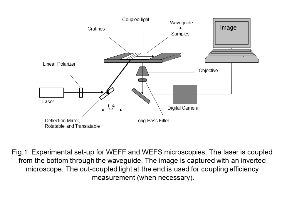
Imaging in the evanescent filed can either be based on fluoresence photons or on scattered photons. Therfore two microscopy technologies are available: WEFF and WEFS microscopy. Fig.1 shows the experimental setup.
WEFF Microscopy
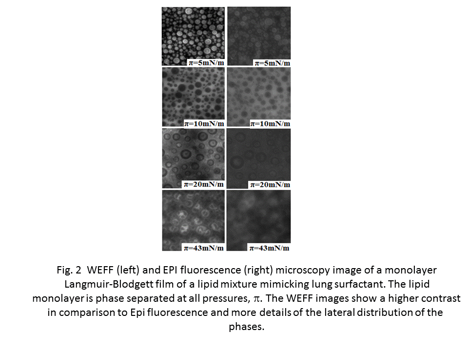
In WEFF microscopy the sample on the waveguide needs to be stained with a fluorophore that can be exited by the wavelength of light propagating in the waveguide. Fig.2 shows an example of WEFF images taken from a phase separted LB film.
Because waveguides allow to propagate light in more than one mode various images illuminated by different modes can be taken. This allows to calculate the distance of the fluorophore from the waveguide surface, thus allowing to plot dye-distance maps. Fig.3 shows an example from an osteblast with a stained plasma membrane.
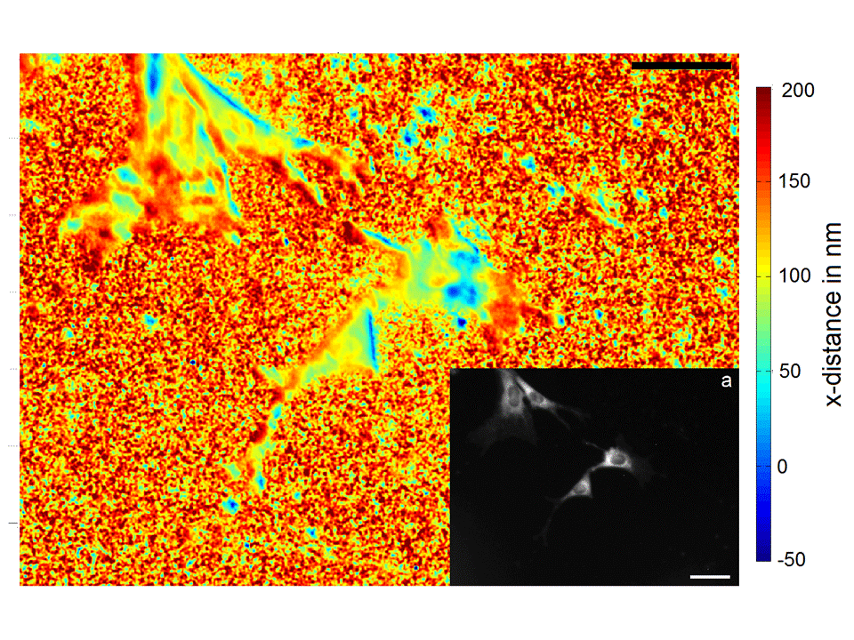
Fig. 3 Dye distance map of an osteoblast cultured on a waveguide. The inset is an overexposed WEFF image which yields the same information as EPI fluorescence microscopy. The dark blue areas are adhesions sites of the cell.
WEFS Microscopy
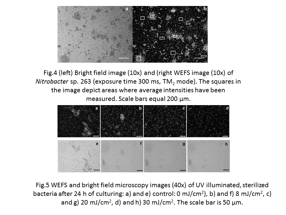
In WEFS microscopy now staining is necessary. The scattered photons out of the evanescent field are used to form an image. An example from biofilms is shown in Fig.4.
Wavguide Absorption Spectroscopy
On the other hand, by coupling white light into a waveguide device and monitoring the out coupled spectrum one should yield information about the absorption of anything located in the evanescent field. The unique possibility to use either TE or TM modes offers an independent insight in in-lane and out-of-plane absorptions independently. For the white light an adequate coupling mechanism has to be used. The outcoupled intensity is be analysed by means of a spectrometer.


